CIESC Journal ›› 2020, Vol. 71 ›› Issue (10): 4808-4819.DOI: 10.11949/0438-1157.20200780
• Material science and engineering, nanotechnology • Previous Articles Next Articles
Xiaojing LI( ),Wen SUN,Yao KANG,Jiangli FAN(),Xiaojun PENG
Received:2020-06-19
Revised:2020-07-23
Online:2020-10-05
Published:2020-10-05
Contact:
Jiangli FAN
李晓静(),孙文,康垚,樊江莉(),彭孝军
通讯作者:
樊江莉
作者简介:李晓静(1991—),女,博士研究生,基金资助:CLC Number:
Xiaojing LI, Wen SUN, Yao KANG, Jiangli FAN, Xiaojun PENG. Synthesis of PEGylation hydroxyapatite drug delivery system and its dual channels fluorescence imaging[J]. CIESC Journal, 2020, 71(10): 4808-4819.
李晓静, 孙文, 康垚, 樊江莉, 彭孝军. PEG化羟基磷灰石纳米体系的制备及双通道荧光成像[J]. 化工学报, 2020, 71(10): 4808-4819.
Add to citation manager EndNote|Ris|BibTeX
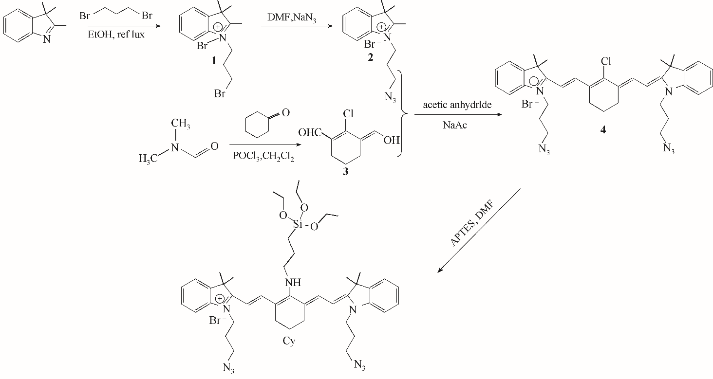
Fig.1 Reaction equation of cyanine dyes and PEG alkynyl
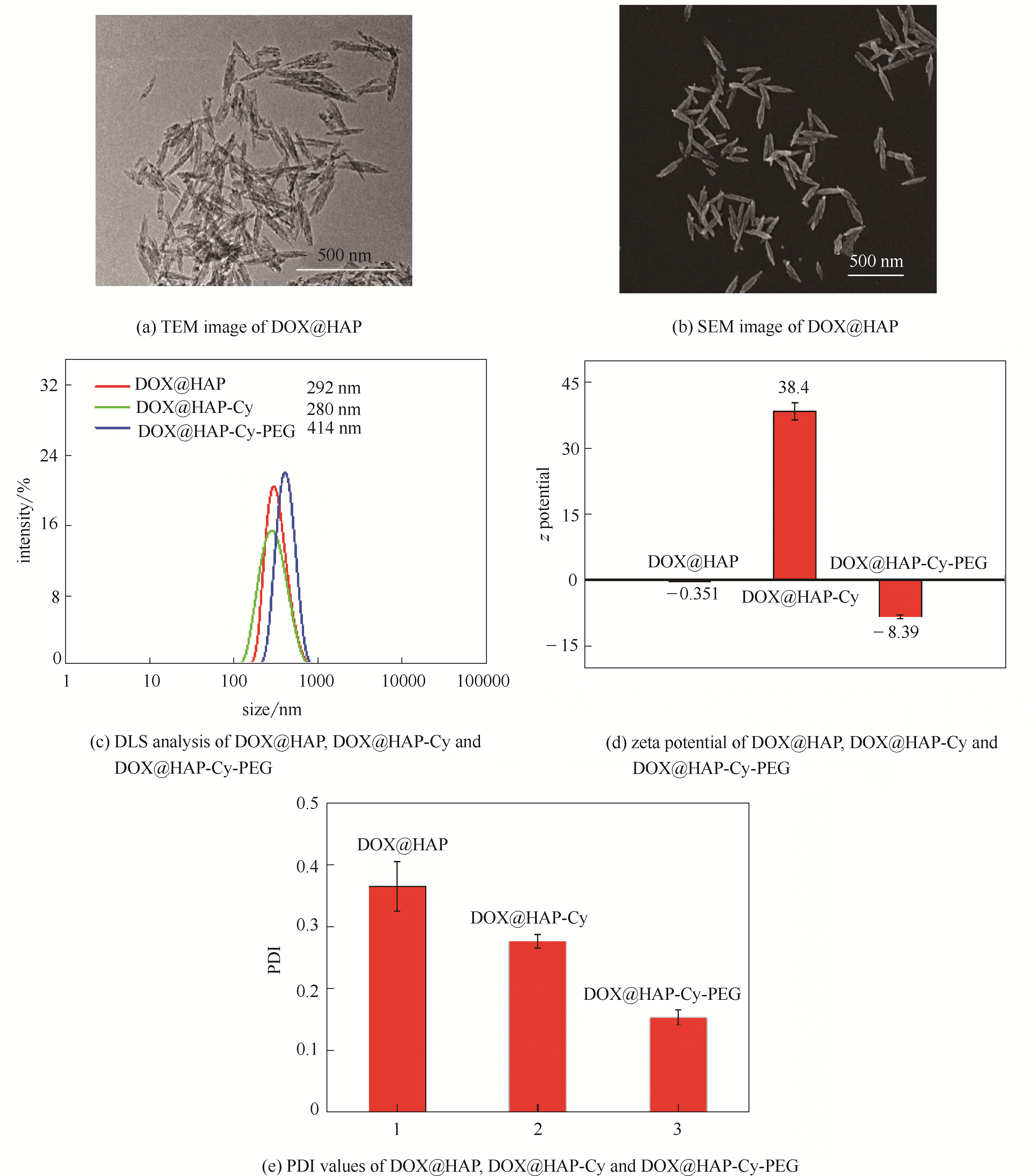
Fig.2 TEM (a) and SEM (b) images of DOX@HAP. DLS analysis (c), zeta potential (d) and PDI values (e) of DOX@HAP,DOX@HAP-Cy and DOX@HAP-Cy-PEG
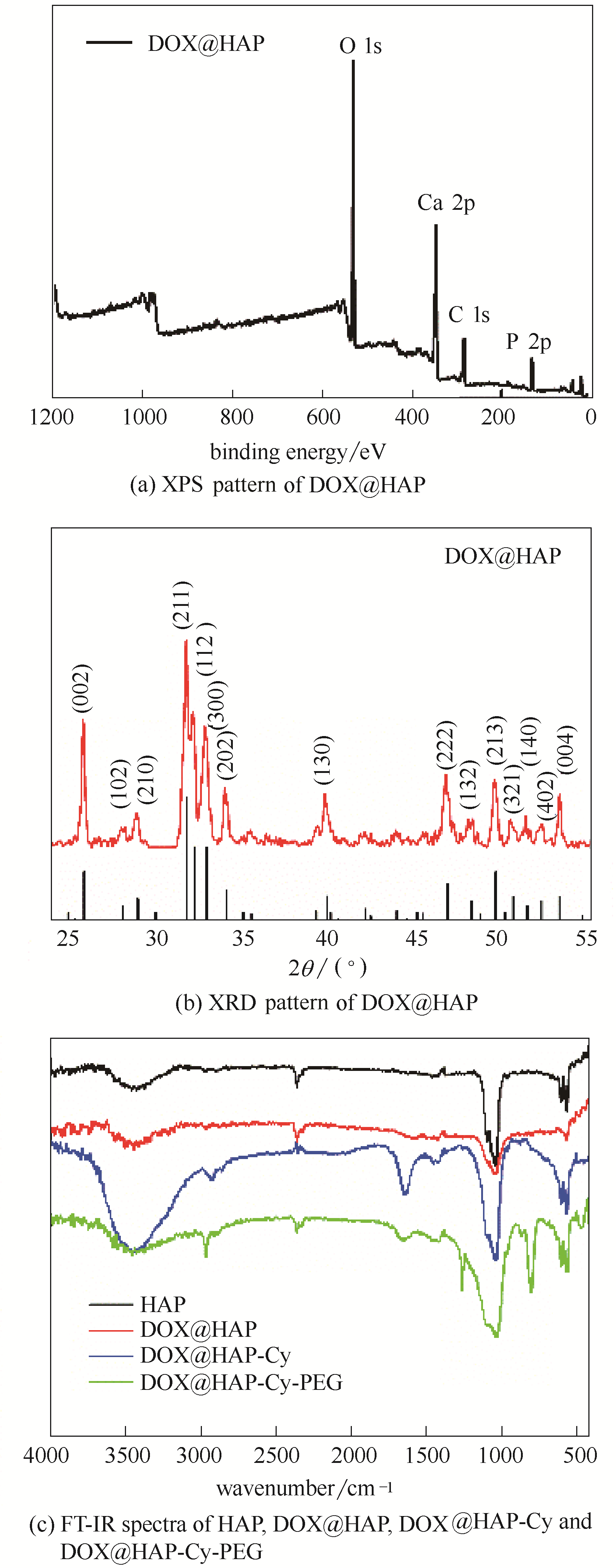
Fig.3 XPS (a) and XRD (b) patterns of DOX@HAP. FTIR spectra (c) of HAP,DOX@HAP,DOX@HAP-Cy and DOX@HAP-Cy-PEG
Fig.4 The absorption spectra (a) and fluorescence spectra (b) of DOX,Cy and DOX@HAP-Cy-PEG
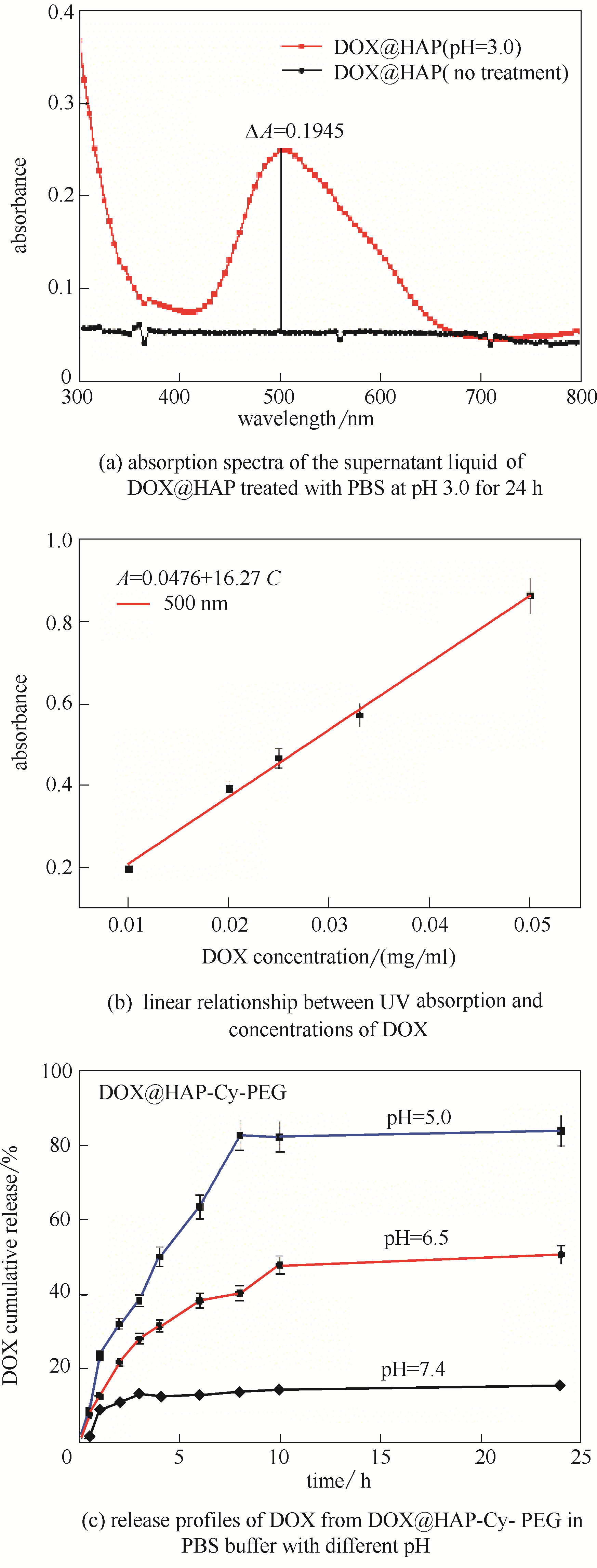
Fig.5 The absorption spectra of the supernatant liquid of DOX@HAP treated with PBS at pH 3.0 for 24 h(a). The linear relationship between UV absorption (λab = 500 nm) and concentrations of DOX: (A = 16.27C + 0.0476)(b). Release profiles of DOX from DOX@HAP-Cy-PEG in PBS buffer with different pH (5.0,6.5,7.4) in 24 h(c)
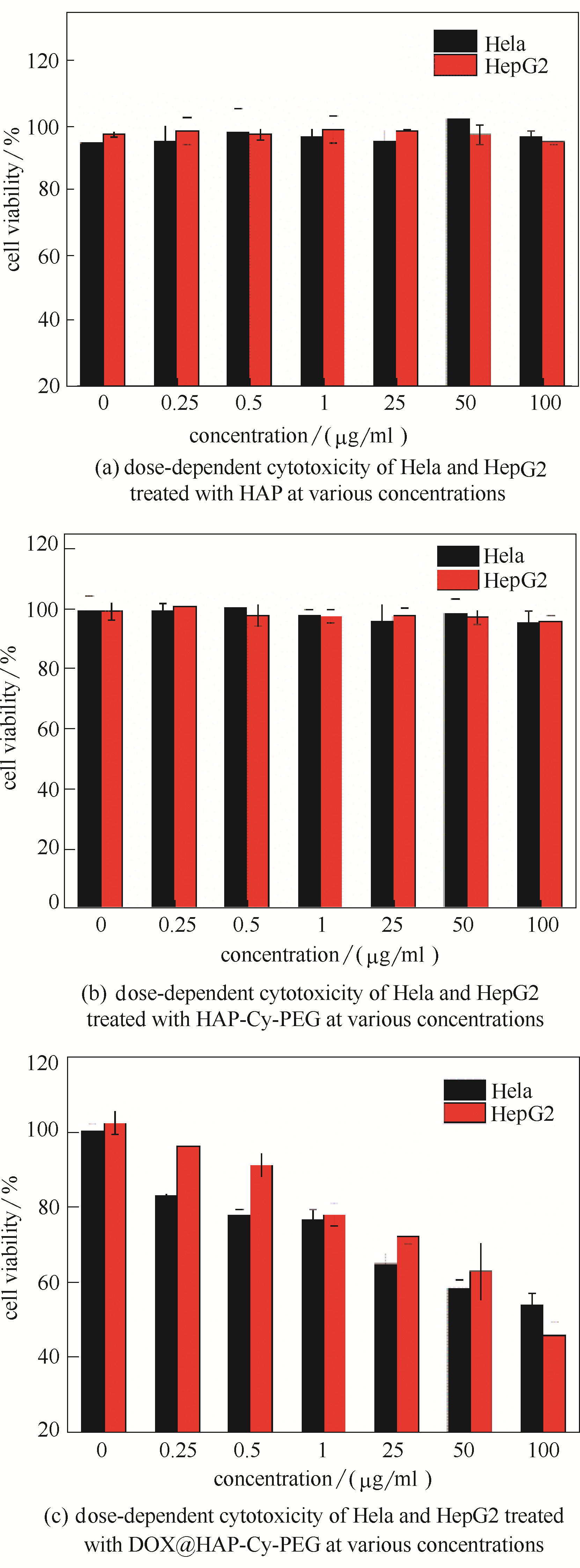
Fig.6 Dose-dependent cytotoxicity of Hela and HepG2 treated with HAP (a),HAP-Cy-PEG (b) and DOX@HAP-Cy-PEG (c) at various concentrations
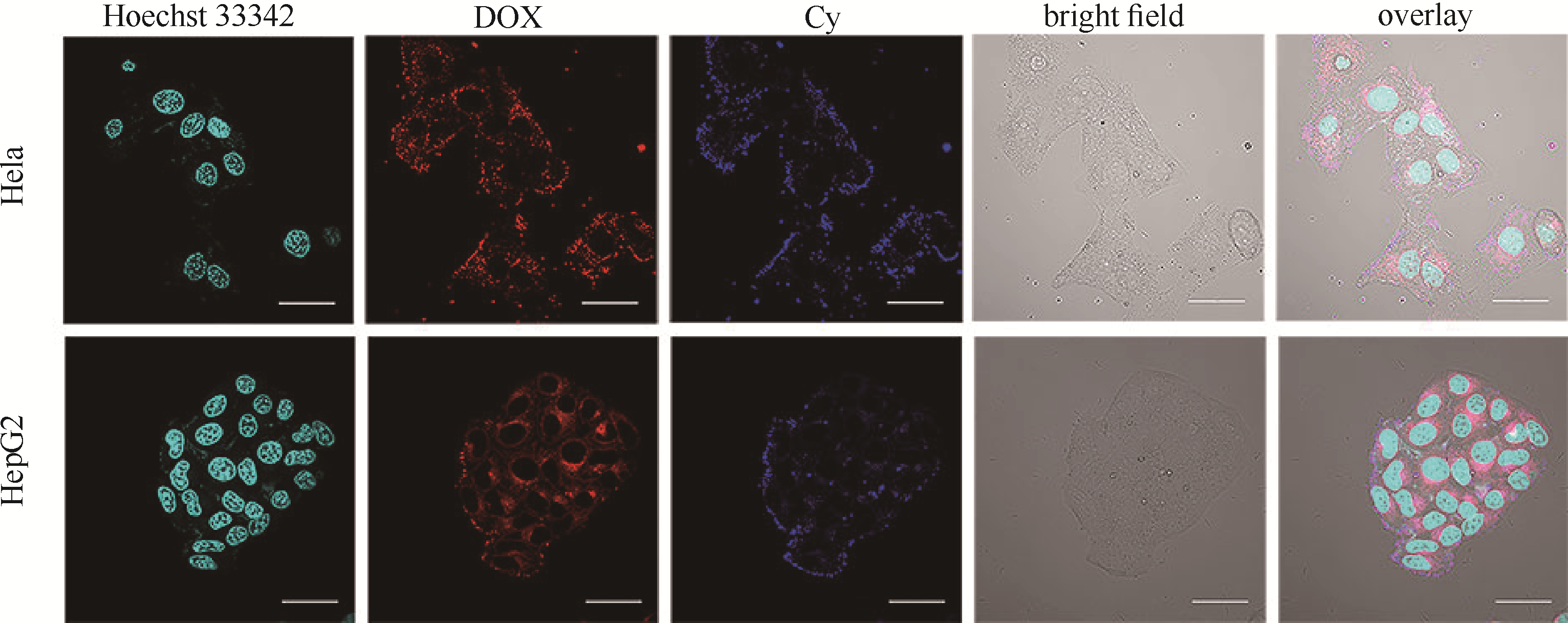
Fig.7 CLSM images of Hela and HepG2 cells incubated with DOX@HAP-Cy-PEG for 4 h by dual channels fluorescence imaging
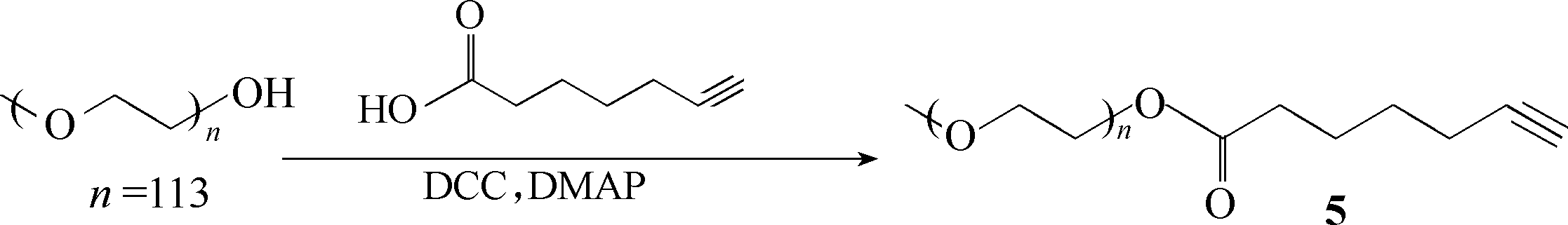
Fig.A1 Synthetic route of PEG-alkyne
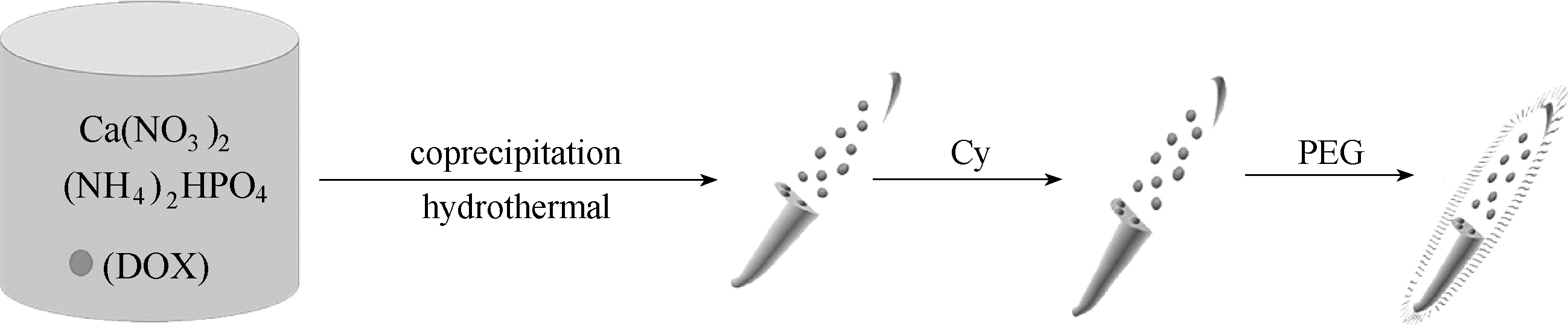
Fig.A2 Synthetic route of DOX@HAP-Cy-PEG
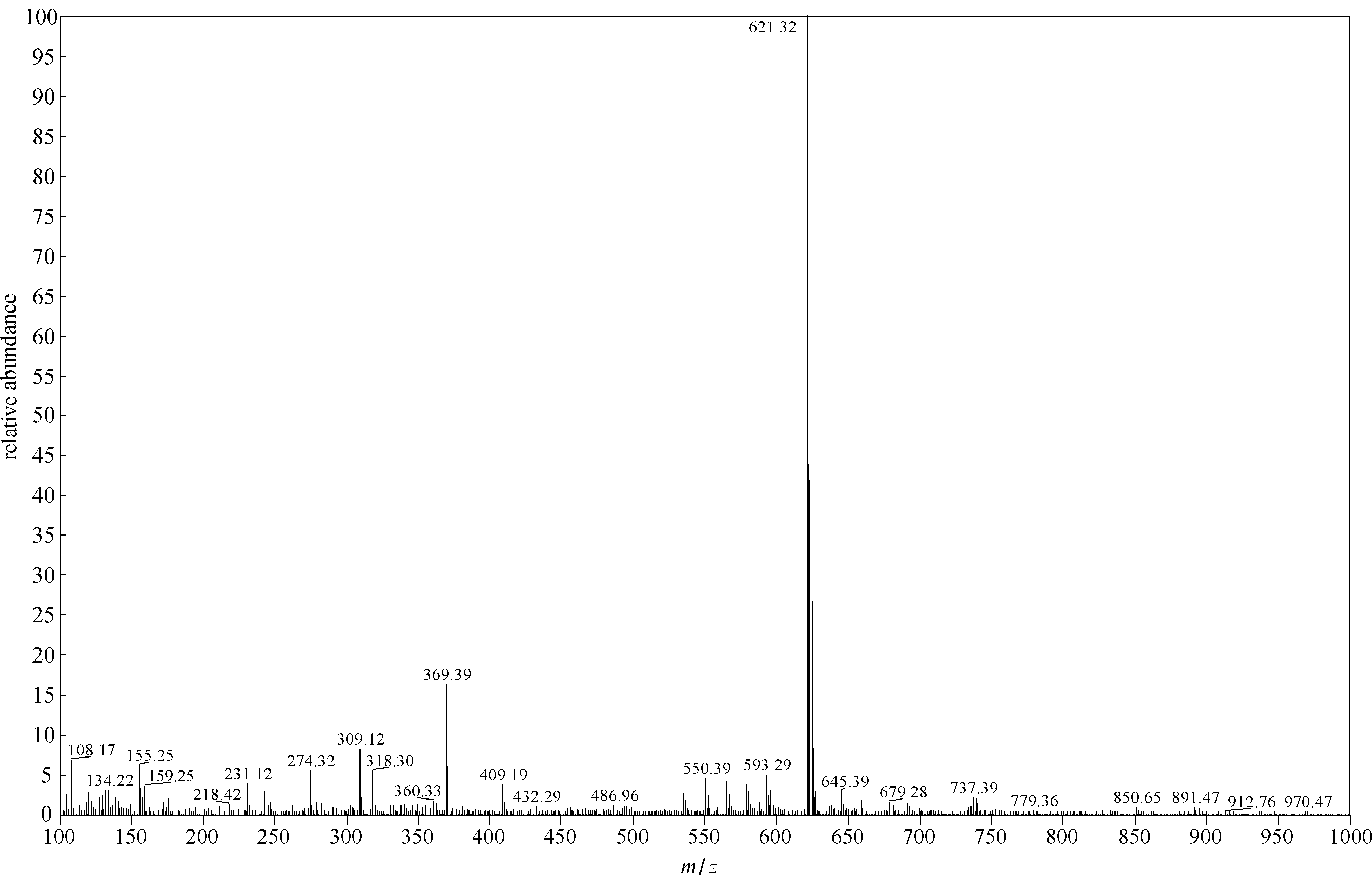
Fig.A3 ESI-MS spectrum of compound 4
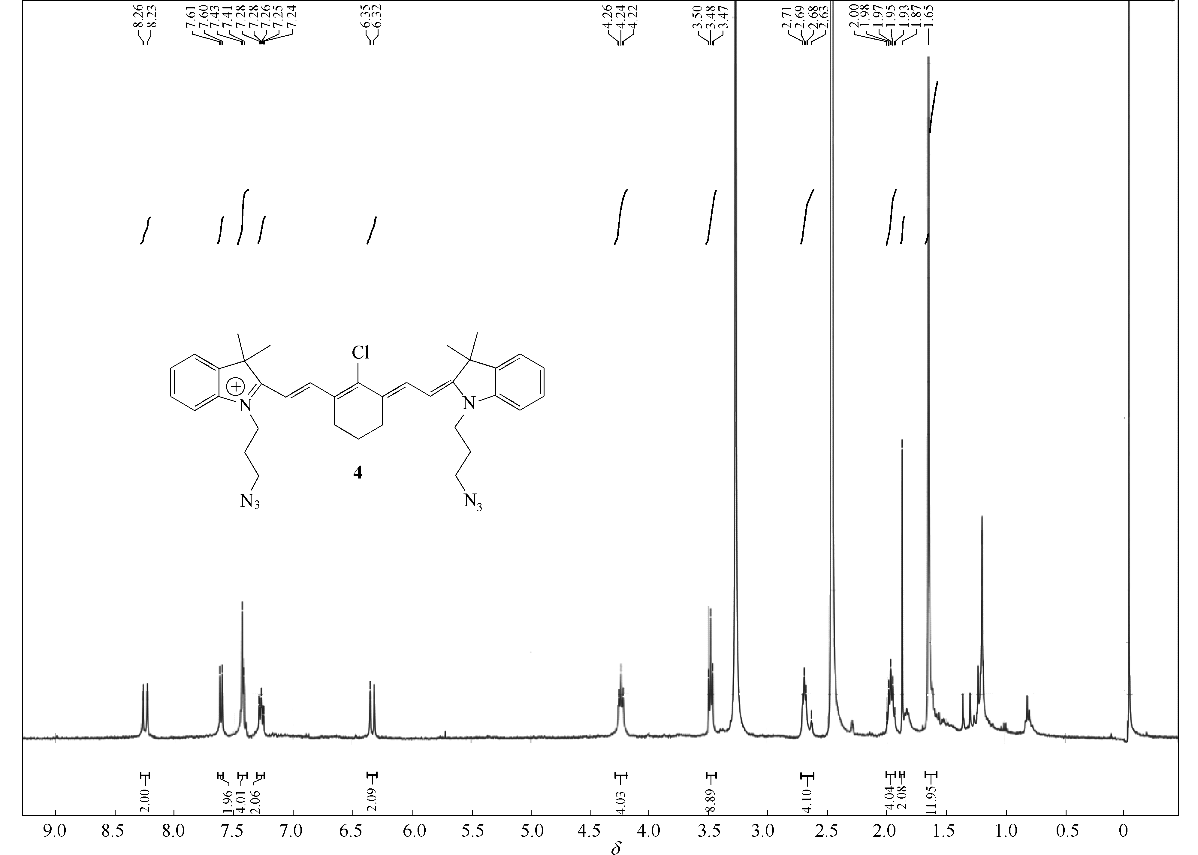
Fig.A4 1H NMR spectrum of compound 4
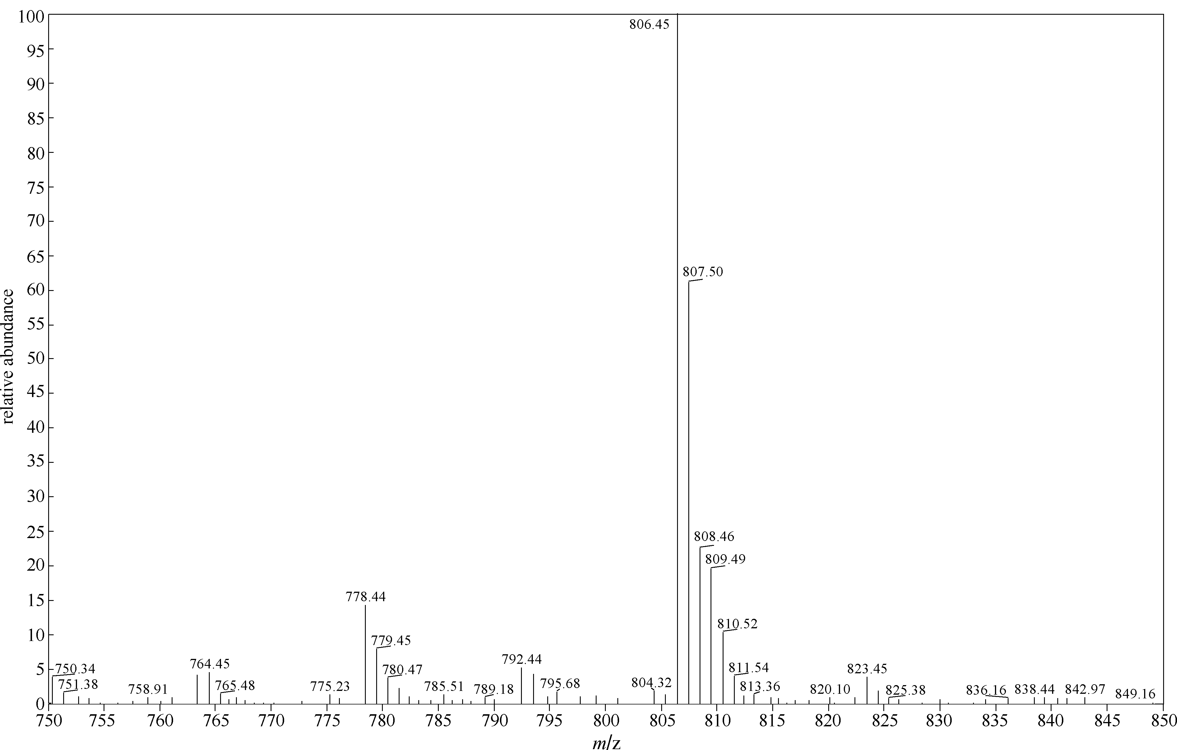
Fig.A5 ESI-MS spectrum of Cy

Fig.A6 1H NMR spectrum of Cy
| Element | Peak binding energy/eV | Content/%(atom) |
|---|---|---|
| P | 132.75 | 8.96 |
| Ca | 346.88 | 14.6 |
| C | 284.49 | 27.75 |
| O | 530.89 | 48.7 |
Table A1 XPS elemental analysis of DOX@HAP
| Element | Peak binding energy/eV | Content/%(atom) |
|---|---|---|
| P | 132.75 | 8.96 |
| Ca | 346.88 | 14.6 |
| C | 284.49 | 27.75 |
| O | 530.89 | 48.7 |
| 1 | Maia A L C, Ferreira C A, Barros A L B, et al. Vincristine-loaded hydroxyapatite nanoparticles as a potential delivery system for bone cancer therapy[J]. J. Drug Target., 2018, 26(7): 592-603. |
| 2 | Kang Y, Sun W, Fan J, et al. Ratiometric real-time monitoring of hydroxyapatite–doxorubicin nanotheranostic agents for on-demand tumor targeted chemotherapy[J]. Mater. Chem. Front., 2018, 2(10): 1791-1798. |
| 3 | Salamanna F, Giavaresi G, Parrilli A, et al. Antiresorptive properties of strontium substituted and alendronate functionalized hydroxyapatite nanocrystals in an ovariectomized rat spinal arthrodesis model[J]. Mater. Sci. Eng. C Mater. Biol. Appl., 2019, 95: 355-362. |
| 4 | 康垚, 王素真, 樊江莉, 等. 无机纳米药物载体在肿瘤诊疗中的研究进展[J]. 化工学报, 2018, 69(1): 128-140. |
| Kang Y, Wang S Z, Fan J L, et al. Progress in inorganic nanomedicine carriers for tumor diagnosis and treatments[J]. CIESC Journal, 2018, 69(1): 128-140 | |
| 5 | Jiang F, Wang D P, Ye S, et al. Strontium-substituted, luminescent and mesoporous hydroxyapatite microspheres for sustained drug release[J]. J Mater. Sci. Mater. Med., 2014, 25(2): 391-400. |
| 6 | Xu Y J, Dong L, Lu Y, et al. Magnetic hydroxyapatite nanoworms for magnetic resonance diagnosis of acute hepatic injury[J]. Nanoscale, 2016, 8(3): 1684-1690. |
| 7 | Abbasi Aval N, Pirayesh Islamian J, Hatamian M, et al. Doxorubicin loaded large-pore mesoporous hydroxyapatite coated superparamagnetic Fe3O4 nanoparticles for cancer treatment[J]. Int. J. Pharm., 2016, 509(1/2): 159-167. |
| 8 | Govindan B, Swarna Latha B, Nagamony P, et al. Designed synthesis of nanostructured magnetic hydroxyapatite based drug nanocarrier for anti-cancer drug delivery toward the treatment of human epidermoid carcinoma[J]. Nanomaterials, 2017, 7(6): 138-154. |
| 9 | Yang Y H, Liu C H, Liang Y H, et al. Hollow mesoporous hydroxyapatite nanoparticles (hmHANPs) with enhanced drug loading and pH-responsive release properties for intracellular drug delivery[J]. J. Mater. Chem. B, 2013, 1(19): 2447-2450. |
| 10 | Sarkar C, Chowdhuri A R, Kumar A, et al. One pot synthesis of carbon dots decorated carboxymethyl cellulose-hydroxyapatite nanocomposite for drug delivery, tissue engineering and Fe3+ ion sensing[J]. Carbohydr. Polym., 2018, 181: 710-718. |
| 11 | Sun W, Fan J, Wang S, et al. Biodegradable drug-loaded hydroxyapatite nanotherapeutic agent for targeted drug release in tumors[J]. ACS Appl. Mater. Interfaces, 2018, 10(9): 7832-7840. |
| 12 | Xiong H, Du S, Ni J, et al. Mitochondria and nuclei dual-targeted heterogeneous hydroxyapatite nanoparticles for enhancing therapeutic efficacy of doxorubicin[J]. Biomaterials, 2016, 94: 70-83. |
| 13 | Pan L, Liu J, He Q, et al. MSN-mediated sequential vascular-to-cell nuclear-targeted drug delivery for efficient tumor regression[J]. Adv. Mater., 2014, 26(39): 6742-6748. |
| 14 | Wu J B, Shi C, Chu G C, et al. Near-infrared fluorescence heptamethine carbocyanine dyes mediate imaging and targeted drug delivery for human brain tumor[J]. Biomaterials, 2015, 67: 1-10. |
| 15 | Gorka A P, Nani R R, Zhu J, et al. A near-IR uncaging strategy based on cyanine photochemistry[J]. J. Am. Chem. Soc., 2014, 136(40): 14153-14159. |
| 16 | Nani R R, Gorka A P, Nagaya T, et al. Near-IR light-mediated cleavage of antibody-drug conjugates using cyanine photocages[J]. Angew. Chem. Int. Ed., 2015, 54(46): 1-5. |
| 17 | Gorka A P, Yamamoto T, Zhu J, et al. Cyanine photocages enable spatial control of inducible cre-mediated recombination[J]. Chembiochem, 2018, 19(12): 1239-1243. |
| 18 | Ma K, Sai H, Wiesner U. Ultrasmall sub-10 nm near-infrared fluorescent mesoporous silica nanoparticles[J]. J. Am. Chem. Soc., 2012, 134(32): 13180-13183. |
| 19 | Nissinen T, Nakki S, Laakso H, et al. Tailored dual PEGylation of inorganic porous nanocarriers for extremely long blood circulation in vivo[J]. ACS Appl. Mater. Interfaces, 2016, 8(48): 32723-32731. |
| 20 | Zhao L, Yuan W, Ang C Y, et al. Silica-polymer hybrid with self-assembled PEG corona excreted rapidly via a hepatobiliary route[J]. Adv. Funct. Mater., 2016, 26(18): 3036-3047. |
| 21 | Su Y Y, Teng Z, Yao H, et al. A Multifunctional PB@mSiO2-PEG/DOX nanoplatform for combined photothermal- chemotherapy of tumor[J]. ACS Appl. Mater. Interfaces, 2016, 8(27): 17038-17046. |
| 22 | Ren T B, Xia W J, Dong H Q, et al. Sheddable micelles based on disulfide-linked hybrid PEG-polypeptide copolymer for intracellular drug delivery[J]. Polymer, 2011, 52(16): 3580-3586. |
| 23 | Ni R, Zhu J, Xu Z, et al. A self-assembled pH/enzyme dual-responsive prodrug with PEG deshielding for multidrug-resistant tumor therapy[J]. J. Mater. Chem. B, 2020, 8(6): 1290-1301. |
| 24 | Selli D, Motta S, Di Valentin C. Impact of surface curvature, grafting density and solvent type on the PEGylation of titanium dioxide nanoparticles[J]. J. Colloid Interface Sci., 2019, 555: 519-531. |
| 25 | Zhou M, Huang H, Wang D, et al. Light-triggered PEGylation/dePEGylation of the nanocarriers for enhanced tumor penetration [J]. Nano Lett., 2019, 19(6): 3671-3675. |
| 26 | Joh D Y, Zimmers Z, Avlani M, et al. Architectural modification of conformal PEG-bottlebrush coatings minimizes anti-PEG antigenicity while preserving stealth properties[J]. Adv. Healthc. Mater., 2019, 8(8): 1801177-1801190. |
| 27 | Hammarson M, Andersson J, Li S, et al. Molecular AND-logic for dually controlled activation of a DNA-binding spiropyran[J]. Chem. Commun., 2010, 46(38): 7130-7132. |
| 28 | Kumar K, Sagar S, Esau L, et al. Synthesis of novel 1H-1,2,3-triazole tethered C-5 substituted uracil-isatin conjugates and their cytotoxic evaluation[J]. Eur. J. Med. Chem., 2012, 58: 153-159. |
| 29 | Kang N Y, Park S J, Ang X W, et al. A macrophage uptaking near-infrared chemical probe CDnir7 for in vivo imaging of inflammation[J]. Chem. Commun., 2014, 50: 6589-6591. |
| 30 | Li B, Lu L, Zhao M, et al. An efficient 1064 nm NIR-II excitation fluorescent molecular dye for deep-tissue high-resolution dynamic bioimaging[J]. Angew. Chem. Int. Ed., 2018, 57(25): 7483-7487. |
| 31 | Chen G, Song F, Wang J, et al. FRET spectral unmixing: a ratiometric fluorescent nanoprobe for hypochlorite[J]. Chem. Commun., 2012, 48: 2949-2951. |
| 32 | Sun W, Parowatkin M, Steffen W, et al. Ruthenium-containing block copolymer assemblies: red-light-responsive metallopolymers with tunable nanostructures for enhanced cellular uptake and anticancer phototherapy[J]. Adv. Healthc. Mater., 2016, 5(4): 467-473. |
| 33 | Zhen X, Zhang J, Huang J, et al. Macrotheranostic probe with disease-activated near-infrared fluorescence, photoacoustic, and photothermal signals for imaging-guided therapy[J]. Angew. Chem. Int. Ed., 2018, 57(26): 7804-7808. |
| 34 | Ha S W, Park J, Habib M M, et al. Nano-hydroxyapatite stimulation of gene expression requires Fgf receptor, phosphate transporter, and Erk1/2 signaling[J]. ACS Appl. Mater. Interfaces, 2017, 9(45): 39185-39196. |
| 35 | 任欣, 金蜀鄂, 李玉宝, 等. 纳米羟基磷灰石增强聚己内酯/明胶纤维膜的制备及其性能[J]. 化工进展, 2020, 39(4): 1439-1446. |
| Ren X, Jin S E, Li Y B, et al. Preparation and performance of nano-hydroxyapatite reinforced polycaprolactone/gelatin fibrous membrane for guided tissue regeneration[J]. Chem. Ind. & Eng. Pro., 2020, 39(4): 1439-1446. |
| [1] | YU Fuqiang, DU Jianjun, LU Yang, MA He, FAN Jiangli, SUN Wen, LONG Saran, PENG Xiaojun. Fabrication of serum albumin-copper phthalocyanine nanoparticles for mitochondria-targeted phototherapy [J]. CIESC Journal, 2021, 72(1): 597-608. |
| [2] | YOU Donghui, CHENG Zhiliang, LI Gan, ZHANG Feng, LYU Fanglei, JIANG Guangbin, QUAN Xuejun, WEI Jianwei, YANG Lu, LI Shuo. Preparation and catalytic degradation property research of novel phthalocyanine [J]. CIESC Journal, 2018, 69(12): 5090-5099. |
| [3] | XU Yanming, ZHAO Ming, LI Jian, REN Qiang, WANG Chenyi. Visible near-infrared amino phthalocyanine-titanium dioxide photocatalyst: preparation and performance [J]. CIESC Journal, 2016, 67(5): 1915-1921. |
| [4] | ZHANG Juan, HU Yanhui, REN Tengjie, LI Weikang, ZHAO Dishun. Photocatalytic oxidation desulfurization by iron phthalocyanine supported on Ti-MCM-41 [J]. CIESC Journal, 2015, 66(9): 3437-3443. |
| [5] | ZHAO Ming, LI Jian, JI Junling, REN Qiang, WANG Chenyi, AI Baolin. Synthesis and application of soluble near-infrared absorption amino phthalocyanine dyes [J]. CIESC Journal, 2015, 66(4): 1577-1584. |
| [6] | ZHANG Juan, REN Tengjie, HU Yanhui, LI Junpan, WANG Chunfang, ZHAO Dishun. Catalytic performance of metal phthalocyanine loaded on MCM-41 molecular sieve in oxidation desulfurization [J]. CIESC Journal, 2014, 65(8): 3012-3018. |
| [7] | LIU Guijin, WANG Hongdi, JIANG Yanbin. Research progress of Zein as carrier for drug delivery systems [J]. CIESC Journal, 2013, 64(10): 3493-3504. |
| [8] | WU Qingxi, YAO Shanjing. Oral colon-specific drug delivery system and its preparation [J]. CIESC Journal, 2013, 64(1): 210-222. |
| [9] | LI Wenfeng, LIU Guijin, LI Lixian, WU Juan, Lü Yangxiao, JIANG Yanbin. Effect of process parameters on co-precipitation of paclitaxel and poly(L-lactic acid) by supercritical antisolvent process [J]. Chin.J.Chem.Eng., 2012, 20(4): 803-813. |
| [10] | NING Zhang-Lei, CHANG Zhi-Dong, LI Wen-Jun, SUN Chang-Yan, ZHANG Jing-Hua, LIU Yang- . Solvothermal synthesis and optical performance of one-dimensional strontium hydroxyapatite nanorod [J]. , 2012, 20(1): 89-94. |
| [11] | SHEN Juan,JIN Bo,JIANG Qiying,HU Yamin,ZHONG Guoqing,HUO Jichuan. Preparation,properties and applications of hydroxyapatite/synthetic polymer composites [J]. , 2011, 30(8): 1749-. |
| [12] | WEN Xin,AN Shengjun. Progress of growth factors containing wound dressing [J]. , 2009, 28(8): 1435-. |
| [13] | SHI Yan,NING Ping,WAND Xueqian,JIANG Ming. Treatment of H2S by modified activated carbon [J]. , 2009, 28(5): 890-. |
| [14] | ZHAO Xuhui, YANG Lingfang, ZUO Yu, XIONG Jinping. Hydroxyapatite Coatings on Titanium Prepared by Electrodeposition in a Modified Simulated Body Fluid [J]. , 2009, 17(4): 667-671. |
| [15] | ZHANG Dan. Application of membrane filter press in pigment industry [J]. , 2009, 28(2): 342-. |
| Viewed | ||||||
|
Full text |
|
|||||
|
Abstract |
|
|||||