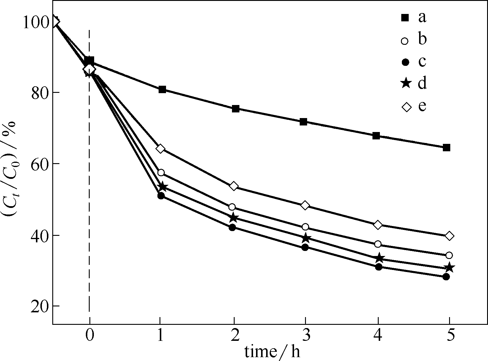
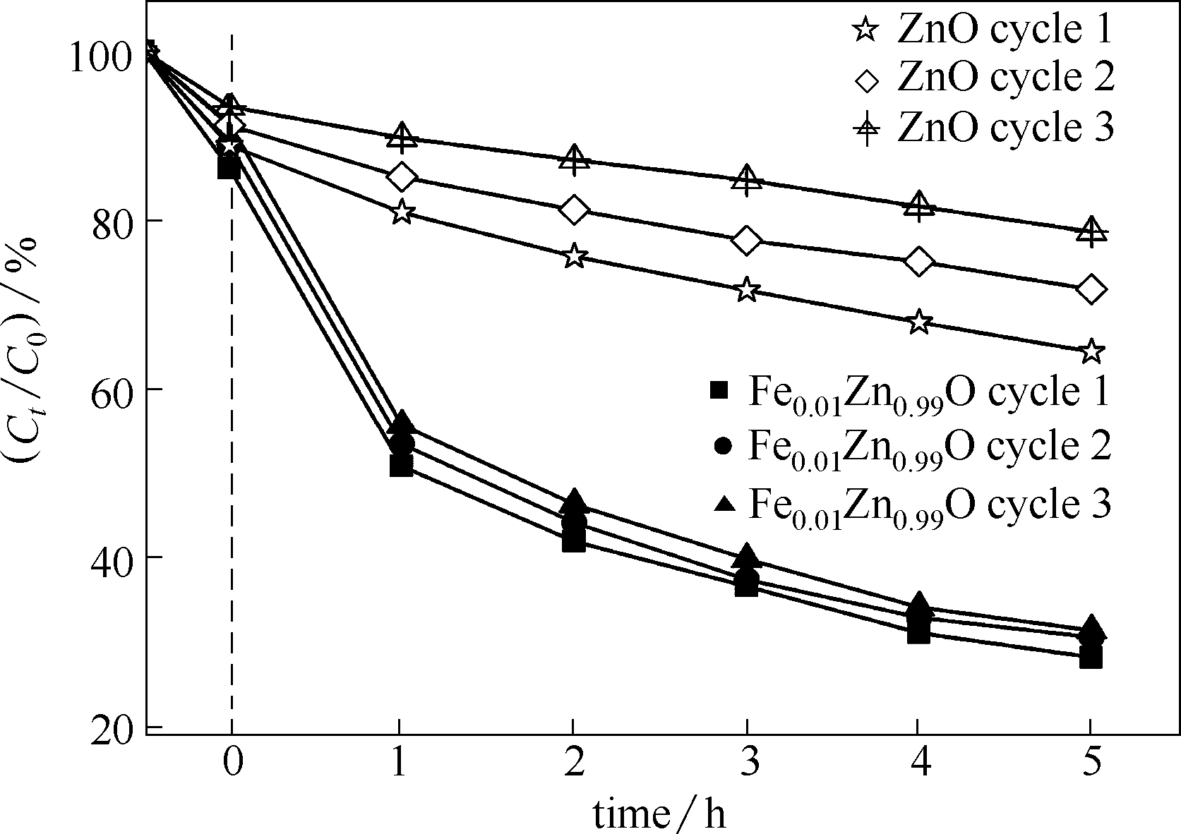
| 1 |
FortunaoE, BarquinhaP, PimentelA, et al. Recent advances in ZnO transparent thin film transistors[J]. Thin Solid Films, 2005, 487(1/2): 205-211.
|
| 2 |
SalehR, DjajaN F. UV light photocatalytic degradation of organic dyes with Fe-doped ZnO nanoparticles[J]. Superlattices and Microstructures, 2014, 74: 217-233.
|
| 3 |
WangY, ZhaoX, DuanL, et al. Structure, luminescence and photocatalytic activity of Mg-doped ZnO nanoparticles prepared by auto combustion method[J]. Materials Science in Semiconductor Processing, 2015, 29: 372-379.
|
| 4 |
丁艳, 马歌, 李良超, 等. M2+(M= Cu, Cd, Ag, Fe) 掺杂氧化锌纳米粉晶的抗菌性能[J]. 无机化学学报, 2014, 30(2): 293-302.
|
|
DingY, MaG, LiL C, et al. Antibacterial activities of doped ZnO nano-powder with M2+ (M=Cu, Cd, Ag and Fe) [J]. Chinese Journal of Inorganic Chemistry, 2014, 30(2): 293-302.
|
| 5 |
KumarR, UmarA, KumarG, et al. Ce-doped ZnO nanoparticles for efficient photocatalytic degradation of direct red-23 dye[J]. Ceramics International, 2015, 41(6): 7773-7782.
|
| 6 |
ShuklaS K, AgorkuE S, MittalH, et al. Synthesis, characterization and photoluminescence properties of Ce 3+-doped ZnO-nanophosphors[J]. Chemical Papers, 2014, 68(2): 217-222.
|
| 7 |
IsmailI M I, AslamM, AlmeelbiT, et al. Ce3+ impregnated ZnO: a highly efficient photocatalyst for sunlight mediated mineralization[J]. RSC Advances, 2014, 4(31): 16043-16046.
|
| 8 |
SharmaD K, SharmaK K, KumarV, et al. Effect of Ce doping on the structural, optical and magnetic properties of ZnO nanoparticles[J]. Journal of Materials Science: Materials in Electronics, 2016, 27(10): 10330-10335.
|
| 9 |
MayJ W, MaJ, BadaevaE, et al. Effect of excited-state structural relaxation on midgap excitations in Co2+-doped ZnO quantum dots[J]. The Journal of Physical Chemistry C, 2014, 118(24): 13152-13156.
|
| 10 |
KumarS, SongT K, GautamS, et al. Structural, magnetic and electronic structure properties of Co doped ZnO nanoparticles[J]. Materials Research Bulletin, 2015, 66: 76-82.
|
| 11 |
RazaW, HaqueM M, MuneerM. Synthesis of visible light driven ZnO: characterization and photocatalytic performance[J]. Applied Surface Science, 2014, 322: 215-224.
|
| 12 |
KanevaN, BojinovaA, PapazovaK, et al. Photocatalytic purification of dye contaminated sea water by lanthanide (La3+, Ce3+, Eu3+) modified ZnO[J]. Catalysis Today, 2015, 252: 113-119.
|
| 13 |
ChenC, MaW, ZhaoJ. Semiconductor-mediated photodegradation of pollutants under visible-light irradiation[J]. Chem. Soc. Rev., 2010, 39: 4206-4219.
|
| 14 |
BaikJ M, KangT W, LeeJ L. Effects of N2O plasma treatment on magnetic properties of (Zn, Mn)O nanorods[J]. Nanotechnology, 2007, 18(9): 095703.
|
| 15 |
翟英娇, 李金华, 陈新影, 等. 镉掺杂氧化锌纳米花的制备及其光催化活性[J]. 中国光学, 2014, 7(1): 124-130.
|
|
ZhaiY J, LiJ H, ChenX Y, et al. Preparation and photocatalytic activity of cadmium-doped zinc oxide nanoflowers[J]. China Optics, 2014, 7(1): 124-130.
|
| 16 |
KolevaM E, AtanasovP A, NedialkovN N, et al. Role of vanadium content in ZnO thin films grown by pulsed laser deposition[J]. Applied Surface Science, 2007, 254(4): 1228-1231.
|
| 17 |
YimazS, ParlakM, ŞÖzcan, et al. Structural, optical and magnetic properties of Cr doped ZnO microrods prepared by spray pyrolysis method[J]. Applied Surface Science, 2011, 257(22): 9293-9298.
|
| 18 |
ShindeV R, GujarT P, LokhandeC D, et al. Mn doped and undoped ZnO films: a comparative structural, optical and electrical properties study[J]. Mater. Chem. Phys., 2006, 96: 326-330.
|
| 19 |
ShishodiaP K. Effect of cobalt doping on ZnO thin films deposited by sol-gel method[J]. Thin Solid Films, 2016, 612: 55-60.
|
| 20 |
ZhaoX, LiuE, RamanujanR V, et al. Effects of rapid thermal annealing on structural, magnetic and optical properties of Ni-doped ZnO thin films[J]. Current Applied Physics, 2012, 12(3): 834-840.
|
| 21 |
HouD L, YeX J, MengH J, et al. Magnetic properties of n-type Cu-doped ZnO thin films[J]. Applied Physics Letters, 2007, 90(14): 142502.
|
| 22 |
SrivastavaA, KumarN, KhareS. Enhancement in UV emission and band gap by Fe doping in ZnO thin films[J]. Opto-Electronics Review, 2014, 22(1): 68-76.
|
| 23 |
YiS S, CuiJ B, LiS, et al. Enhanced visible-light photocatalytic activity of Fe/ZnO for Rhodamine B degradation and its photogenerated charge transfer properties[J]. Applied Surface Science, 2014, 319: 230-236.
|
| 24 |
SalehR, DjajaN F. UV light photocatalytic degradation of organic dyes with Fe-doped ZnO nanoparticles[J]. Superlattices and Microstructures, 2014, 74: 217-233.
|
| 25 |
LmaiF, MoubahR, AmiriE A, et al. Spin wave study and optical properties in Fe-doped ZnO thin films prepared by spray pyrolysis technique[J]. Optical Materials, 2016, 57: 28-33.
|
| 26 |
WuX, WeiZ, ZhangL, et al. Optical and magnetic properties of Fe doped ZnO nanoparticles obtained by hydrothermal synthesis[J]. Journal of Nanomaterials, 2014, 2014: 1-6.
|
| 27 |
LiX, HouM, HanB, et al. Solubility of CO2 in a choline chloride+ urea eutectic mixture[J]. Journal of Chemical & Engineering Data, 2008, 53(2): 548-550.
|
| 28 |
AbbottA P, CullisP M, GibsonM J, et al. Extraction of glycerol from biodiesel into a eutectic based ionic liquid[J]. Green Chemistry, 2007, 9(8): 868-872.
|
| 29 |
PhadtareS B, ShankarlingG S. Halogenation reactions in biodegradable solvent: efficient bromination of substituted 1-aminoanthra-9, 10-quinone in deep eutectic solvent (choline chloride: urea) [J]. Green Chemistry, 2010, 12(3): 458-462.
|
| 30 |
HarishkumarH N, MahadevanK M, MasagalliJ N, et al. Synthesis and fluorescence study of phenylcoumarin / cyanophenylbenzocoumarin-3-carboxylates[J]. Organic Comm-unications, 2012, 5(4): 196-208.
|
| 31 |
PawaP M, JaragK J, ShankarlingG S. Environmentally benign and energy efficient methodology for condensation: an interesting facet to the classical Perkin reaction[J]. Green Chemistry, 2011, 13(8): 2130-2134.
|
| 32 |
SutrisnoA, LiuL, DongJ, et al. Solid-state 91Zr NMR characterization of layered and three-dimensional framework zirconium phosphates[J]. The Journal of Physical Chemistry C, 2012, 116(32): 17070-17081.
|
| 33 |
MaldonadoM, OleksiakM D, ChintaS, et al. Controlling crystal polymorphism in organic-free synthesis of Na-zeolites[J]. Journal of the American Chemical Society, 2013, 135(7): 2641-2652.
|
| 34 |
LiaoH G, JiangY X, ZhouZ Y, et al. Shape-controlled synthesis of gold nanoparticles in deep eutectic solvents for studies of structure-functionality relationships in electrocatalysis[J]. Angewandte Chemie International Edition, 2008, 47(47): 9100-9103.
|
| 35 |
AbbottA P, CapperG, DaviesD L, et al. Selective extraction of metals from mixed oxide matrixes using choline-based ionic liquids[J]. Inorganic Chemistry, 2005, 44(19): 6497-6499.
|
| 36 |
DongJ Y, HsuY J, WongD S H, et al. Growth of ZnO nanostructures with controllable morphology using a facile green antisolvent method[J]. The Journal of Physical Chemistry C, 2010, 114(19): 8867-8872.
|
| 37 |
ShaktiN, GuptaP S. Structural and optical properties of sol-gel prepared ZnO thin film[J]. Applied Physics Research, 2010, 2(1): 19-28.
|
| 38 |
BundesmannC, AshkenovN, SchubertM, et al. Raman scattering in ZnO thin films doped with Fe, Sb, Al, Ga, and Li[J]. Applied Physics Letters, 2003, 83(10): 1974-1976.
|
| 39 |
WangJ B, HuangG J, ZhongX L, et al. Raman scattering and high temperature ferromagnetism of Mn-doped ZnO nanoparticles[J]. Applied Physics Letters, 2006, 88(25): 252502.
|
| 40 |
SinghP, KaushalA, KaurD. Mn-doped ZnO nanocrystalline thin films prepared by ultrasonic spray pyrolysis[J]. Journal of Alloys and Compounds, 2009, 471(1/2): 11-15.
|
| 41 |
KaramatS, RawatR S, LeeP, et al. Structural, elemental, optical and magnetic study of Fe doped ZnO and impurity phase formation[J]. Progress in Natural Science: Materials International, 2014, 24(2): 142-149.
|
| 42 |
RambuA P, NicaV, DobromirM. Influence of Fe-doping on the optical and electrical properties of ZnO films[J]. Superlattices and Microstructures, 2013, 59: 87-96.
|
| 43 |
CuiJ, SunJ, LiuX, et al. Fabrication of hierarchical flower-like porous ZnO nanostructures from layered ZnC2O4·3Zn(OH)2 and gas sensing properties[J]. Applied Surface Science, 2014, 308: 17-23.
|
| 44 |
RajaK, RameshP S, D. StructuralGeetha, FTIR and photoluminescence studies of Fe doped ZnO nanopowder by co-precipitation method[J]. Spectrochimica Acta Part A: Molecular and Biomolecular Spectroscopy, 2014, 131: 183-188.
|
| 45 |
MitraP, MondalS. Structural and morphological characterization of ZnO thin films synthesized by SILAR[J]. Progress in Theoretical and Applied Physics, 2013, 1: 17-31.
|
| 46 |
ParraP A, PeralesP O, SinghalR, et al. Structural, optical, and magnetic characterization of monodisperse Fe-doped ZnO nanocrystals[J]. Journal of Applied Physics, 2008, 103(7): 07D121.
|
| 47 |
李奡麒, 陈玉娟, 胡晓宇, 等. 低共熔溶剂辅助水热法合成分层球状微/纳米 ZnO 晶体及其光催化性能[J]. 高等学校化学学报, 2015, 36(1): 165-170.
|
|
LiW Q, ChenY J, HuX Y, et al. Synthesis of layered spherical micro/nano ZnO crystals by hydrothermal solvent-assisted hydrothermal method[J]. Chemical Journal of Chinese Universities, 2015, 36(1): 165-170.
|
| 48 |
ZhangQ, LiuJ K, WangJ D, et al. Atmospheric self-induction synthesis and enhanced visible light photocatalytic performance of Fe3+ doped Ag-ZnO mesocrystals[J]. Ind. Eng. Chem. Res., 2014, 53:13236-13246
|
 ),徐存英2,3(
 京公网安备 11010102001995号